For the first time in history, a movable iMRI with an ‘integrated seismic system’ has been successfully implemented.
IMRIS Senior Technical Specialist, Jorge Kern, takes us through the seismic challenge, the creative solution, and the revolutionary innovation that’s bringing intraoperative imaging to places it’s never been.
A CHALLENGE BENEATH THE SURFACE.
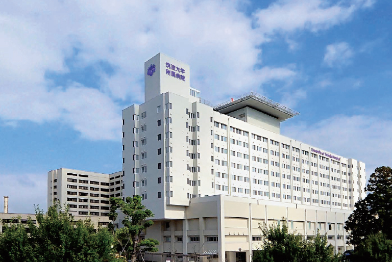
Like many of the world’s leading neurological institutions, The University of Tsukuba Hospital was ready to invest in intraoperative imaging – a technology that enables surgeons to obtain real-time, diagnostic-quality images during operations. For neurosurgeons, introducing this innovation would mean better resection-rates. For patients, it would mean better outcomes. For the University of Tsukuba Hospital, it would mean a new level of institutional prestige.
However, just beneath the surface of the promising opportunity, there was a challenge.
Each year, Japan experiences more than 1,500 earthquakes. That kind of frequent seismic activity can be detrimental to intraoperative imaging: in terms of image-quality, reliability and safety. For the University of Tsukuba Hospital, those factors made intraoperative imaging immensely challenging.
It was up to our team of designers, architects, engineers and clinical specialists to find a solution.
CRAFTING AN INNOVATIVE SOLUTION.
To bring intraoperative imaging to the University of Tsukuba Hospital, we had to accomplish 3 things, despite being in a seismic event-prone region:
- Performance. The slightest vibration can lead to ‘motion artifacts’ in MR images, rendering them unusable for diagnostics. To meet the strict Vibration Isolation Requirement of MRI technology, we engineered a device that, when triggered by a certain amount of vibration, causes MR imaging to stop when image quality is compromised.
- Safety. There are strict regulations for medical technology in earthquake-prone regions. We engineered a solution to meet those regulations – protecting patients and clinical staff – while functioning with existing technology and workflows. The seismic foot, for example, is a device designed to prevent the magnet from disengaging from the rails during an earthquake.
- Reliability. We developed what we call a ‘passive solution’, rather than ‘active’. It couldn’t be a sensor that is affected by seismic activity because the sensor could potentially fail. The solution had to be something ingrained in the structure itself that would stop the magnet if an earthquake occurred.
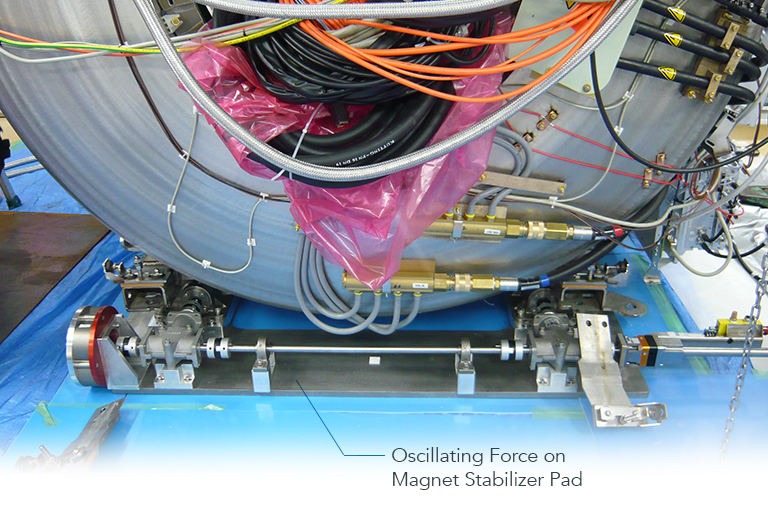
The ‘seismic stabilizer system’ we created is the first of its kind.
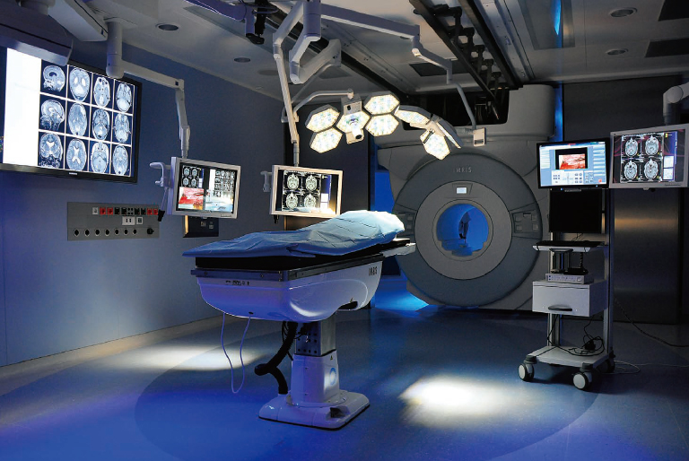
It consists of a stabilizer pad, a bolt-on accessory mounted directly under the magnet. During the operation, as the iMRI travels into the IMRIS Surgical Theatre, the pad is elevated about 25 mm above the ground. Then, when it’s time for imaging, the pad lowers to the floor. The hanging rails support the actual weight of the magnet, while the stabilizer prevents it from swinging side-to-side during seismic activity.
That means guaranteed performance, safety, and stability.
Next, it was time to put our innovation to the test.
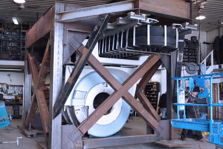
We brought the seismic stabilizer system to the University of Buffalo’s Structural Engineering and Earthquake Simulation Laboratory, where it underwent rigorous testing on a ‘shaker table’. By simulating the conditions of a powerful earthquake, we ensured the device would function as predicted.
Now, it was time to introduce it to the real world.
ELEVATING THE HUMAN EXPERIENCE.
Since the installation, the seismic stabilizer system has been an overwhelming success.
Ninety-one patients have undergone neurosurgical intervention using the University of Tsukuba Hospital’s IMRIS Surgical Theatre. During that time, there have been 169 seismic events in the area, and the system continues to function as it was designed.
IMPACTING GLOBAL HEALTHCARE.

The seismic stabilizer system is a tremendous achievement for the University of Tsukuba Hospital.
But it’s also an achievement for patients, surgeons and institutions around the world.
Right now, hospitals everywhere are demanding the intraoperative imaging capabilities of the IMRIS Surgical Theatre, and some of them are located in seismic event-prone regions like Japan and California. Now, with this new technology, we can bring better outcomes to their institution – wherever they are.
– Jorge Kern, IMRIS Senior Technical Specialist